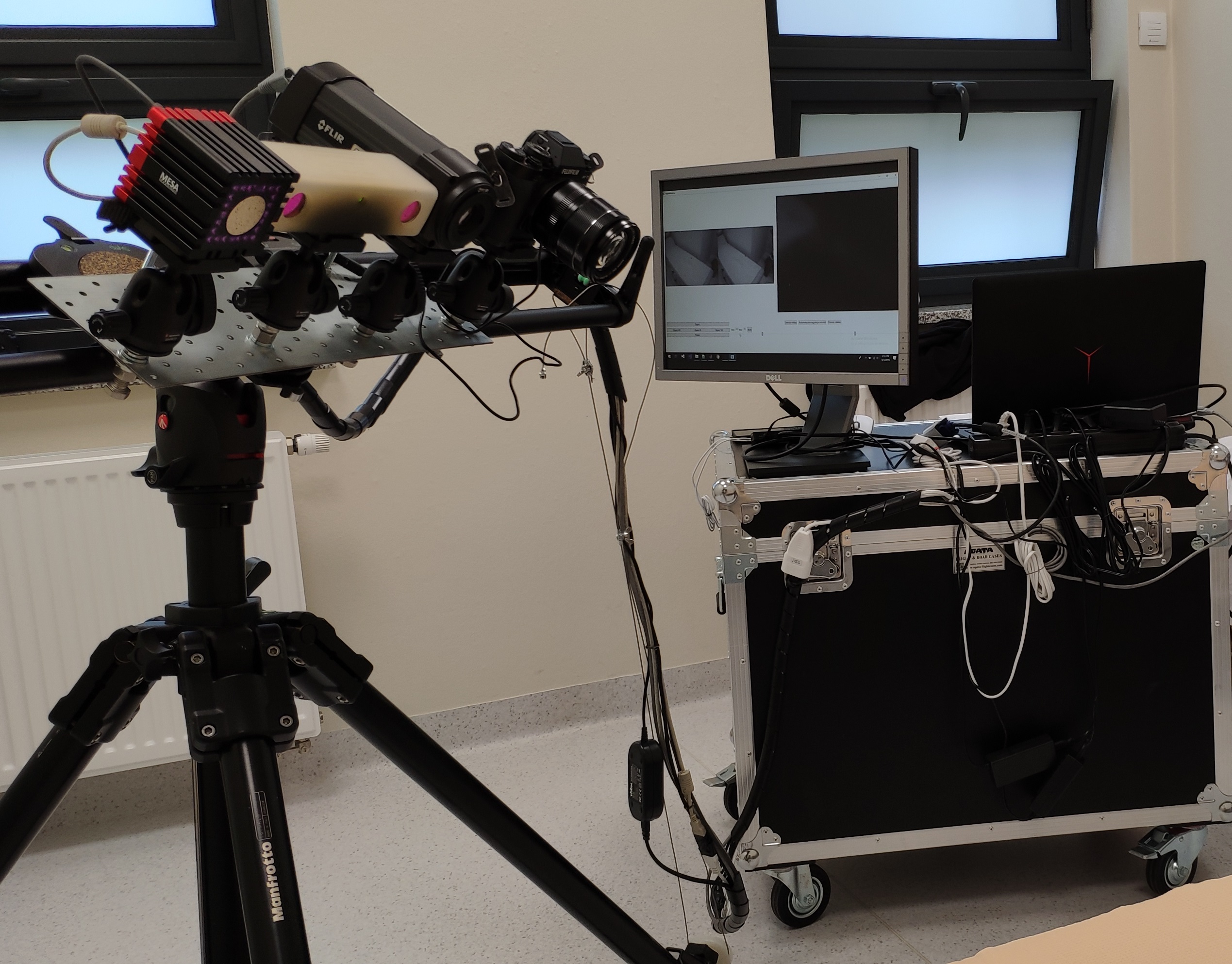
Acquisition system
Cameras used
Wound images are recorded using a set of four cameras. The image data include color photography, thermal imaging, stereovision and depth perception. All the cameras are mounted on a slider with their optical axes parallel. The slider movement enables the cameras to capture data at the same angle once the slider is correctly positioned. Images are taken sequentially after setting the slider in a correct position for each camera.

Color image
Wound color 2D image is acquired at 1920x1080 resolution using a FujiFilm X-T1 digital camera.
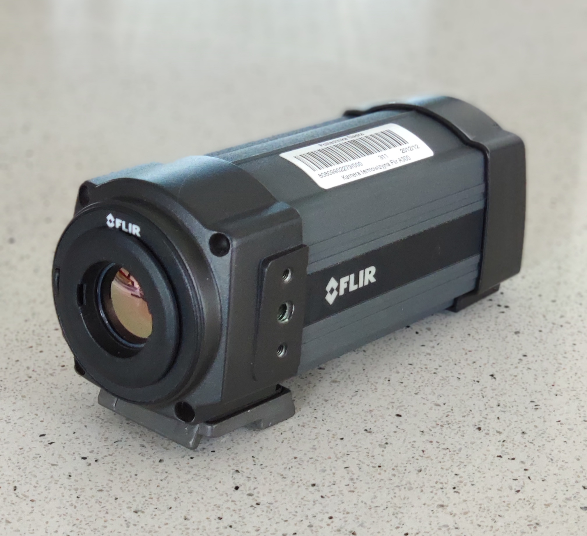
Thermal image
The used FLIR A300 thermal camera captures 2D wound thermal map at 320x240 resolution.

Stereovision
The MicronTracker Hx40 is used to capture the scene (left and right lens saved separately) together with the reconstructed 3D data (point cloud).

Depth perception
A Time-of-Flight camera, SwissRanges SR4000, is used to obtain the depth data of the scene. Several frames at 176x144 resolution are captured in order to choose a single frame or take average of frames.
Data acquisition protocol
Two sets of image data are recorded - calibration and wound data. During the calibration phase a checkerboard is used in the place of the patient. A checkerboard image allows the spatial correspondence between images from all four cameras to be found and is used to derive the similarity transforms. In the meantime medical history is collected and the patient is prepared for the study. After the calibration is over, the patient is positioned, the wound dressing is taken off and the wound data is recorded in sequence: thermal image, stereovision (left and right images), color photography and finally the depth data. If required - the procedure is repeated at another viewing angle. New wound dressing is made before the patient leaves. After the data is acquired, an experienced surgeon performs manual delineation of the wound area in the color image and prepares a case report. The acquired calibration data enables further processing of the wound images - their alignment and fusion giving a multimodal view of the wound.