Data organization
The database is divided into three separate datasets: unprocessed data, processed data and cases descriptions.
Unprocessed data
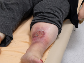
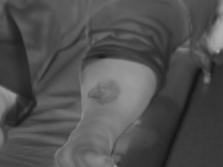
The first part contains the acquired image data from four modalities: color camera (FujiFilm X-T1), thermal camera (FLIR A300), stereo camera (MicronTracker Hx40) and depth camera (SwissRanger SR4000) organized in the following structure of folders case_X/day_Y/[calib|data]/scene_Z/ where case_X is unique patient identifier, day_Y contains data acquired at Yth day from the start of the recordings and calib and data folders contain the images organized in scenes. If the wound had to be examined from several angles several scenes are present. Each scene contains the images of the wound acquired by each of the four cameras. Calibration data are images of a test pattern (checkerboard) required to properly align the images of the wound. For the details of the image acquisition please see the Acquisition system.
Processed data
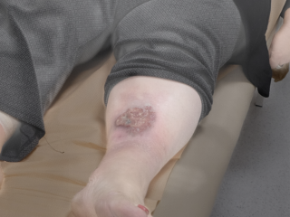
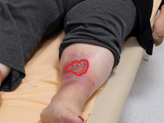
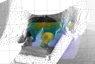
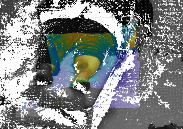
The second part contains the wound images after the processing. The folder structure resembles the tree of the unprocessed data: case_X\day_Y\results\scene_Z. Images avaialble there are aligned (registered) and fused/textured to obtain a multi-modality presentation of the wound. Also the used spatial transformation details are available.
Cases descriptions
The third part of the database contains additional data about the patient (age, sex, etc), the applied wound treatment, paths and indicators to the available image data.