Multi-Modal Image Acquisition Stand for Chronic Wounds Monitoring and Visualization
Motivation
Chronic wound therapy monitoring is commonly carried out using a five-point Torrance or Wagner scale (in Poland). The only measurement performed is the diameter of the wound. Photographic documentation is also common. Due to the increasing trend of chronic wounds, there is a need to develop new tools that will allow monitoring of the progress and treatment of chronic skin wounds to be done effectively.
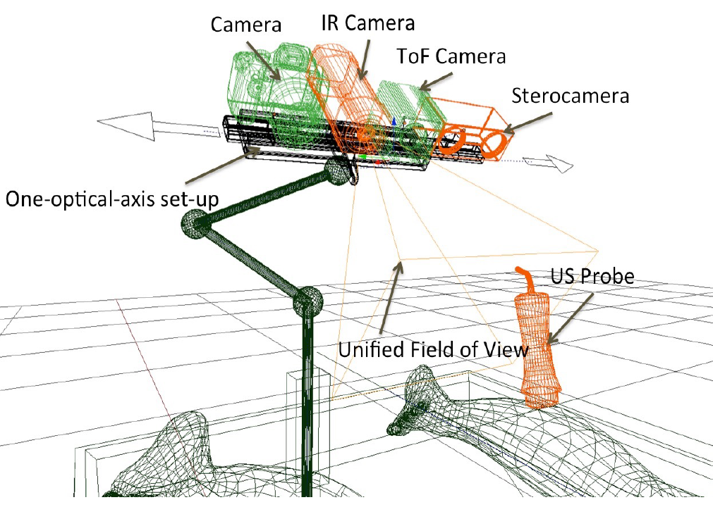
Fig. 1. Concept of a measurement station for the three-dimensional wound model generation.
Concept
The concept of chronic wounds monitoring by generating a three-dimensional wound model involves using image data from the measuring stand (Fig. 1) which is composed of:
- color camera (Fujifilm X-T1, Fujifilm Holdings Corp., Japan) operating at 1920x1080 full HD image resolution,
- stereoscopic camera and optical tool tracking system (MicronTracker Hx40, ClaroNav Inc., Canada) operating at 1024x768 image resolution, thermal imaging camera (FLIR A300, FLIR Systems Inc., USA) operating at 320x240 image resolution,
- and Time-of-Flight camera (SwissRanger SR4000, Mesa Imaging AG, Switzerland) operating at 176x144 image resolution.
In addition, as an extension of the above system, there is a possibility to include an ultrasound image for visualization of deep changes in the tissues within the wound.
The stand calibration procedure allows fusion in a common coordinate system all the image data obtained from the cameras listed. Figure 2 shows the principle:
- three-dimensional geometric model of the wound surface obtained from the sterovision and/or depth perception camera,
- wound thermal image used to texture the 3D wound model,
- wound color image used to texture the 3D wound model,
- possibility to integrate a US image presenting the cross-section of the wound.
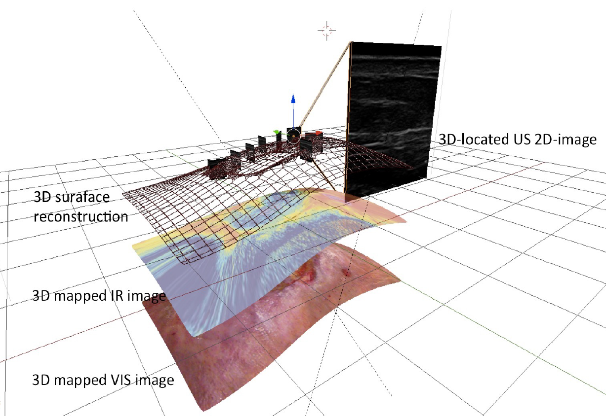
Fig. 2. Fusion of the acquired image data from various sources into a single multimodal 3D model of the wound.
Further work
The use of multimodal image fusion makes it possible to visualize the degree of blood supply to the wound, including the spatial location of its individual areas. Thus it enables the development of a method for monitoring the wound healing process and the selection of parameters objectivizing the description of the state of the wound. The miniaturization of the developed measuring stand and preparation of a visualization software would allow the system to be implemented for general use.
Funding
The prototype of the measuring stand was prepared and tested as part of the grant supported by the Polish National Science Centre (Narodowe Centrum nauki) grant No.: UMO-2016/21/B/ST7/02236.